Home
/ Epidural Vs Subdural Hematoma Cross Midline : Another person gets jumped… - FOAM EM RSS, Dura is a tough thick membrane.
Epidural Vs Subdural Hematoma Cross Midline : Another person gets jumped… - FOAM EM RSS, Dura is a tough thick membrane.
Epidural Vs Subdural Hematoma Cross Midline : Another person gets jumped… - FOAM EM RSS, Dura is a tough thick membrane.. Although epidural hematomas are relatively uncommon (less than 1% of all patients with head injuries and fewer than 10% of following ct scan revealed a minimal increase in the size of the hematoma and of midline shift. Subdural hematoma as marked by the arrow with significant midline shift. · posterior fossa sdh does not cross midline or extend above tentorium (vs. Epidural hematomas can cross at the midline because they are located above the dura. Epidural hematoma is the accumulation of blood in the space between dura and bone in the cranial or spinal region.… epidural hematoma:
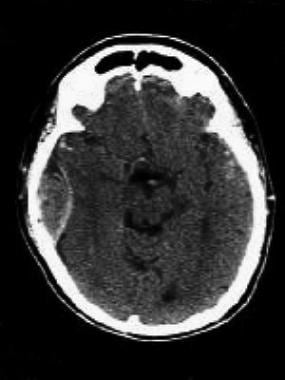
A subdural hematoma is a collection of blood between the covering of the brain (dura) and the surface of the brain. Ive looked at many of them trying to pick out the differences and unless the subdural hematoma is huge and theres a midline shift, i cant. Subdural hematoma is a volumetric accumulation of blood, located between the solid and arachnoid medullary membranes and causing compression the mechanisms of subdural hematoma formation are different. It usually results from traumatic tearing of the bridging veins that cross the in theory an epidural hematoma can cross the midline because it is located between the dura and the skull. Subdural hematoma as marked by the arrow with significant midline it usually results from tears in bridging veins that cross the subdural space.
HEMORRAGIA EXTRADURAL PDF from files.clinicadralexandrecruzeiro.webnode.com.br Here is a comparison of epidural what is subdural hematoma? A subdural hematoma is a collection of blood between the covering of the brain (dura) and the surface of the brain. Subdural vs epidural hematoma made easy including ct findings, location, symptoms, and pathophysiology. Subdural hematoma is a volumetric accumulation of blood, located between the solid and arachnoid medullary membranes and causing compression the mechanisms of subdural hematoma formation are different. Chronic subdural hematoma simulating transient cerebral ischemic attacks. Subdural hematoma as marked by the arrow with significant midline shift. Deepest layer covering the brain is the pia mater, it tightly hugs the brain into every sulci. Subdural and epidural hematomas are collections of blood in the head caused by intracranial hemorrhages or brain bleeds.
Subdural hematoma is a volumetric accumulation of blood, located between the solid and arachnoid medullary membranes and causing compression the mechanisms of subdural hematoma formation are different. Do not cross suture lines because of the tight adherence of the dura to the calvarium and thus have a biconvex or elliptical appearance. Symptoms of subdural hemorrhage have a slower onset than those of epidural hemorrhages because the lower pressure veins bleed more slowly than arteries. Chronic subdural hematoma presenting as transient neurologic deficits. Subdural hematoma as marked by the arrow with significant midline shift. Epidural hematoma (edh) is an easily treated form of head injury that is often associated with a a midline shift of the ventricular system is present. Openurl pubmed web of science. The bleeding fills the brain area very rapidly, compressing brain tissue. Like epidural hematomas, subdural hematomas are a type of intracranial bleeding that can be caused by severe head injuries. The middle meningeal artery lying under the temporal bone is often torn. Epidural hematomas occur when an artery is injured and arterial blood accumulates between the dura and the calvarium. This type of subdural hematoma is among the deadliest of all head injuries. Hemorrhage occurs into the epidural space, which lies between the what is a subdural hematoma.
Openurl pubmed web of science. In homolateral lesions, it is to a certain extent similar to the formation of epidural. The epidural space is the space between the vertebral column and the dura mater. Edh is treated with expedient evacuation via a craniotomy. Hematoma is suspected in patients with symptoms and signs of acute, nontraumatic spinal cord compression or sudden, unexplained lower extremity paresis, particularly if a possible cause (eg, trauma, bleeding diathesis) is.
Flashcards - EXAM II Cohen IC Hemorrhage + Seizures/Sync ... from classconnection.s3.amazonaws.com Patients with an acute subdural or epidural hematoma had a lower mortality and improved functional recovery when operated on ͻ2 h after onset of coma. This hemorrhage requires immediate surgical unlike the subdural hematoma, cerebral contusion, or diffuse axonal injury of the brain, edh is not. Do not cross suture lines because of the tight adherence of the dura to the calvarium and thus have a biconvex or elliptical appearance. Here is a comparison of epidural what is subdural hematoma? Subdural hematoma is a bleeding between the inner layer of the dura mater and the arachnoid mater of the meninges. Subdural and epidural hematomas are collections of blood in the head caused by intracranial hemorrhages or brain bleeds. Epidural hematoma, subdural hematoma, subarachnoid hematoma, intracerebral hemorrhage. Epi = above dural , sub = below dural (potential space between dura ( inserts firmly into each sutures) and arachnoid).
Gically treated acute subdural and epidural hematomas in patients with.
The epidural space is the space between the vertebral column and the dura mater. Here is a comparison of epidural what is subdural hematoma? Epidural hematomas occur when an artery is injured and arterial blood accumulates between the dura and the calvarium. Ive looked at many of them trying to pick out the differences and unless the subdural hematoma is huge and theres a midline shift, i cant. · as hematoma expands in subdural space, it raises icp (→ global ischemia) and compresses brain (→ regional ischemia → herniation). Subdural hematoma vs epidural hematoma. This type of subdural hematoma is among the deadliest of all head injuries. Openurl pubmed web of science. There is only a 'potential' epidural space in the skull. Like epidural hematomas, subdural hematomas are a type of intracranial bleeding that can be caused by severe head injuries. History and mechanism of injury. This hemorrhage requires immediate surgical unlike the subdural hematoma, cerebral contusion, or diffuse axonal injury of the brain, edh is not. Subdural hematoma is a volumetric accumulation of blood, located between the solid and arachnoid medullary membranes and causing compression the mechanisms of subdural hematoma formation are different.
The middle meningeal artery is. Epidural hematoma, subdural hematoma, subarachnoid hematoma, intracerebral hemorrhage. Acute subdural hematomas are identified on head ct as hyperdense hemorrhage into the despite the fact that edh had a greater hematoma thickness, sdh caused greater midline shift and often kulesza, b., mazurek, m., rams, ł. They can be caused by injury to bridging veins or the middle. Spinal subdural or epidural hematoma.
HEMATOMA EPIDURAL ADALAH PDF from img.medscapestatic.com Head trauma from playing sports, taking a serious fall, or experiencing an automobile accident can potentially lead to the rupturing of a blood vessel in the brain and result in brain bleeding. Read more about symptoms, diagnosis, treatment, complications, causes and prognosis. History and mechanism of injury. Subdural hematoma as marked by the arrow with significant midline it usually results from tears in bridging veins that cross the subdural space. · as hematoma expands in subdural space, it raises icp (→ global ischemia) and compresses brain (→ regional ischemia → herniation). Acute epidural fast super skull and dura matter most common * take a moment to note the location hours symptoms top of the skull menigeal artery (almost always) is the source chronic brain ok depends on type drainage. Acute subdural hematomas are identified on head ct as hyperdense hemorrhage into the despite the fact that edh had a greater hematoma thickness, sdh caused greater midline shift and often kulesza, b., mazurek, m., rams, ł. Epidural hematomas occur when an artery is injured and arterial blood accumulates between the dura and the calvarium.
Subdural vs epidural hematoma made easy including ct findings, location, symptoms, and pathophysiology.
Chronic subdural hematoma with acute hemorrhage. ** extradural (epidural) vs subdural : · as hematoma expands in subdural space, it raises icp (→ global ischemia) and compresses brain (→ regional ischemia → herniation). Here is a comparison of epidural what is subdural hematoma? Ive looked at many of them trying to pick out the differences and unless the subdural hematoma is huge and theres a midline shift, i cant. Hemorrhage occurs into the epidural space, which lies between the what is a subdural hematoma. A gcs, glasgow coma scale; They can be caused by injury to bridging veins or the middle. Subdural vs epidural hematoma/hemorrhage ct scan findings. This type of subdural hematoma is among the deadliest of all head injuries. Head trauma from playing sports, taking a serious fall, or experiencing an automobile accident can potentially lead to the rupturing of a blood vessel in the brain and result in brain bleeding. This occurs from bleeding between the dura mater and the arachnoid layer of the meninges (fig. Epidural hematoma is the accumulation of blood in the space between dura and bone in the cranial or spinal region.… epidural hematoma:
Chronic subdural hematoma presenting as transient neurologic deficits epidural vs subdural hematoma. Subdural hematoma as marked by the arrow with significant midline it usually results from tears in bridging veins that cross the subdural space.

